S = Soe−bD
S = Soe−bD
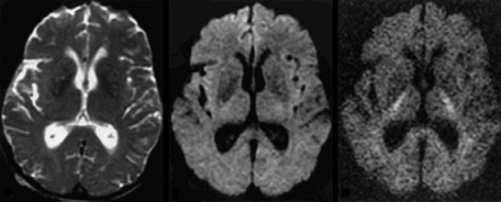
The term e−bD thus behaves very much like the T2-weighting term e−TE/T2 found in many other pulse sequences. The value of b is selected by the operator prior to imaging. This choice controls the degree of observed diffusion-weighting similar to the way choosing TE affects T2-weighting. Diffusion may thus be thought of as another relaxation mechanism in addition to T1 and T2. In pulse sequences without extra diffusion gradients, this relaxation mechanism is relatively unimportant, affecting the final signal by no more than 5%. When diffusion gradients are applied, however, the effects of diffusion are significantly amplified and become the dominant mechanism of tissue contrast.
|
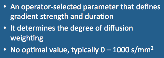
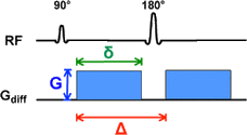
The term "b-value" derives from the landmark 1965 paper by Stejskal and Tanner in which they described their pulsed gradient diffusion method. This technique still forms the basis for most modern DWI pulse sequences and consists of two strong gradient pulses of magnitude (G) and duration (δ), separated by time interval (Δ). The formula for b, specific to this particular implementation only, is shown in the diagram right.
|
Advanced Discussion (show/hide)»
Although the historical Stejskal-Tanner equation for b is given above and found in most textbooks, this particular implementation is seldom used in modern clinical or experimental systems. Rather than pure rectangular pulses, either sinusoidal or trapezoidal pulses are more frequently employed. For sinusoidal pulses the equation becomes b = 4γ²G²δ²(Δ−δ/4)/π². If trapezoidal pulses with rise times ξ are used, then b = γ²G²[δ²(Δ−δ/3) + ξ³/30 −δξ²/6].
Diffusion schemes other than the pulsed gradient method have also been studied, with the calculation of the b-value depending on the details of the gradient scheme used. For example, if a single long diffusion gradient G is applied during the time TE, then b = γ²G²TE³/12. If a fast spin echo technique with n echoes is used, then b = γ²G²TE³/12n².
Burdette JH, Durden DD, Elster AD, Yen YF. High b-value diffusion-weighted MRI of normal brain. J Comput Assist Tomogr 2001; 25:515-519.
Kingsley PB, Monahan WG. Selection of the optimum b factor for diffusion-weighted
magnetic resonance imaging assessment of ischemic stroke. Mag Reson Med 2004; 51:996-1001.
Stejskal EO, Tanner JE. Spin diffusion measurements: spin echoes in the presence of
time-dependent field gradient. J Chem Phys 1965; 42(1):288-292
How do you make a DW image?
In body imaging a starting b-value of 50 (s/mm²) instead of b=0 is often used. Why is this?


